Single-cell RNA sequencing presents an opportunity to deconstruct cellular networks but is limited by the loss of biological information. In this study, a new method of fixation before dissociation using a deep eutectic solvent (DES) is presented.
This method enables multiple domains of in vivo biological data, including morphology, RNA, proteins and posttranslational modifications to be preserved. The study demonstrates how deep eutectic based fixatives will allow the accurate reconstruction of in vivo cellular networks and uncover links between intracellular pathways.
The workflow:
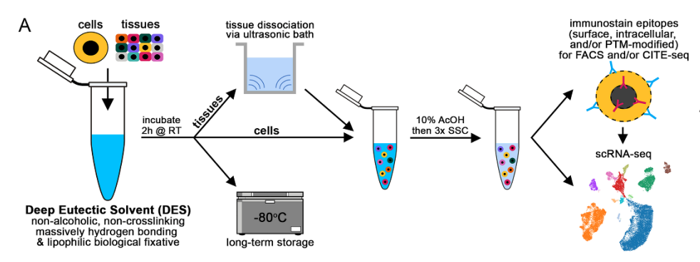
Figure 1. DES fixation allows long-term preservation of RNA and in vivo cell morphology after ultrasonic dissociation. (A) Schematic overview of fixation before dissociation using DES. h, hours. RT, room temperature. SSC, saturated sodium chloride. AcOH, acetic acid. PTM, post-translational modifications. FACS, fluorescence-activated cell sorting.
The result
The study used vivoPHIX™ to rapidly penetrate the sample and fix human retina as well as mouse bone marrow and colon. vivoPHIX™ is included within one subset of DES.
DES are a class of compounds with unique properties for next generation fixatives. vivoPHIX™, rapidly penetrates to fix bacterial, plant and animal cells.
DES fixation allows the long-term stabilization of biomolecules, while weakening cell-to-cell interactions. This facilitates dissociation post-fixation.
This means that DES-based compounds are an attractive alternative to traditional biological fixatives and have the potential to preserve in vivo biology.
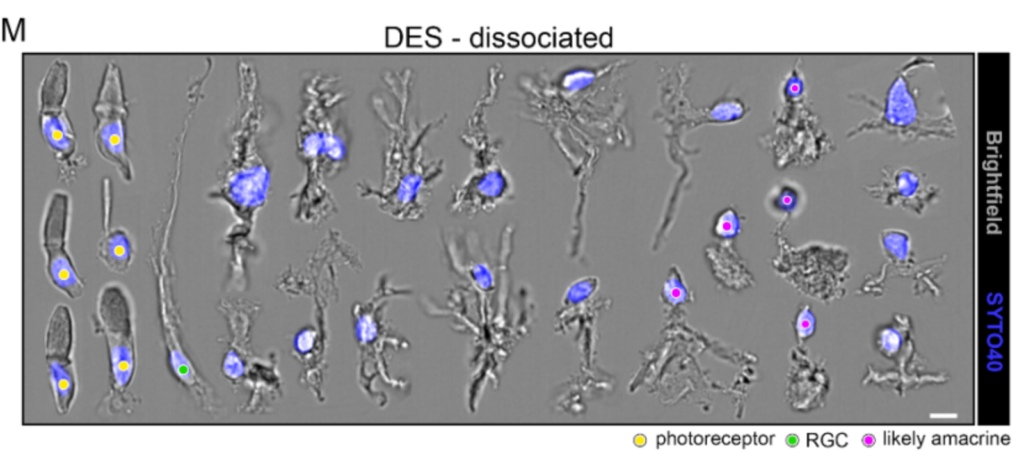
Above: Collage of imaging flow cytometry photos from DES fixed and ultrasonically dissociated human retina from a non-diseased donor. Scale bar=7μm. Yellow circles=photoreceptors, green circle=retinal ganglion cells (RGC), magenta circles=likely amacrine cells. In this figure, you can see the remarkably well preserved cell morphologies (Figure 1M)